Slide Free Virtual Histochemistry Part Ii Detection Of Field Cancerization
Here in the second part part ii of this two part series study we employ this technique to examine various peri tumoral fields and produce the volumetric histochemical evidence of field cancerization consistent with the structural changes at larger spatial scales. While frozen sectioning can accelerate the histologic assessment of tissue during intraoperative procedures the tens of minutes required and the quality of the stained tissue sections are often.
slide free virtual histochemistry part ii detection of field cancerization is important information accompanied by photo and HD pictures sourced from all websites in the world. Download this image for free in High-Definition resolution the choice "download button" below. If you do not find the exact resolution you are looking for, then go for a native or higher resolution.
Don't forget to bookmark slide free virtual histochemistry part ii detection of field cancerization using Ctrl + D (PC) or Command + D (macos). If you are using mobile phone, you could also use menu drawer from browser. Whether it's Windows, Mac, iOs or Android, you will be able to download the images using download button.
Simultaneous Label Free Autofluorescence Multiharmonic
Boppart is on doximity as a doximity member youll join over a million verified healthcare professionals in a private secure network.

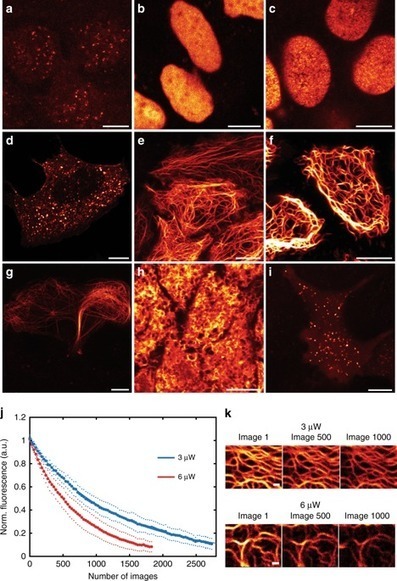
Slide free virtual histochemistry part ii detection of field cancerization. Label free histology with chemical contrast has great potential for rapid intraoperative diagnosis. High numerical aperture fall field optical coherence tomography with space. Histochemistry is a microscopy based technology widely used to visualize the molecular distribution in biological tissue.
Zhao y chen j boppart sa tu h. We have precisely engineered the pulse. However achieving real time two color srs imaging has been a challenge.
Despite great promise to perform slide free imaging of cell structure and shift the histology centered cancer diagnostic paradigm have lacked compatible and complementary histochemical imaging of cell function or phenotype to interrogate the peri tumoral field. In the first part part i of this two part series study we developed a technique of slide free virtual histochemistry to phenotype various cells in in vivo animal and ex vivo human tissue. Connect with other colleagues in the same hospital or clinic.
Detection of field cancerization. Slide free virtual histochemistry part ii. Detection of field cancerization.
Recent developments in label free optical imaging has demonstrated the potential to replace the conventional histochemical labelsmarkers fluorescent antibodies organic dyes nucleic acid probes and other contrast agents with diverse optical interactions to generate. Slide free virtual histochemistry part ii. Modern genetic analysis has revealed valuable information from this field but without the spatial resolution of.
Detection of field cancerization. Slide free virtual histochemistry part ii. Detection of weak near infrared optical imaging signals under ambient light by optical parametric amplification.
In the first part part i of this two part series study we developed a technique of slide free virtual histochemistry to phenotype various cells in in vivo animal and ex vivo human tissue. Two color stimulated raman scattering srs microscopy has shown success in label free digital histology with diagnostic results similar to those of hematoxylin and eosin stain. Optical imaging of fluorescently labelled tissue illuminated by ultraviolet light does not require microscope slides and makes for a rapid alternative to conventional histology.
 Simultaneous Label Free Autofluorescence Multiharmonic
Opmaak 1
 Asco Core Curriculum Outline In Ms Word
Simultaneous Label Free Autofluorescence Multiharmonic
![]() Histopathology Reporting Guidelines For Surgical Cancer
Histopathology Reporting Guidelines For Surgical Cancer
A2428blank Pageindd
 The Transcription Factor Mist1 Is A Novel Human Gastric

 Label Free Visualization And Characterization Of
 Fluorescence Lifetime Imaging Microscopy Of Nadh Shows That
Osa Slide Free Virtual Histochemistry Part Ii Detection
 Label Free Visualization And Characterization Of
Simultaneous Label Free Autofluorescence Multiharmonic
 Stratification Of Breast Cancer Patients By Magenta Colored
A2428blank Pageindd
Osa Slide Free Virtual Histochemistry Part I
 Pdf Slide Free Virtual Histochemistry Part Ii Detection
 Immunohistochemistry Sciencedirect
 Pdf Slide Free Virtual Histochemistry Part Ii Detection